- Visibility 78 Views
- Downloads 10 Downloads
- DOI 10.18231/j.johs.2024.040
-
CrossMark
- Citation
Benign but troublesome: A case study of irritational fibroma in the oral cavity
Introduction
Fibroma is a frequent submucosal reaction to damage from teeth or dental prosthesis. It is sometimes referred to as fibrous polyp or polypus. In 1846, it was originally reported.[1], [2] It arises from local irritation or injury and is also referred to as reactive focal fibrous hyperplasia.[3], [4] It is most frequently found in 1.2% of people and is frequently made up of Type I and Type III collagen.[5] This lesion is the result of irritations like calculus, overhanging edges, trauma, and dental appliances.[6] Three distinct kinds of fibromas exist:
Hard fibroma.
Soft fibroma.
Other fibroma types include angio fibroma, cystic fibroma, myxo fibroma, and cemento-ossifying fibroma. Hard fibroma has numerous fibres and few cells, whereas soft fibroma has many loosely packed cells.[7]
Case Reports
Case 1
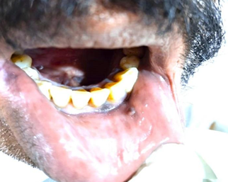
A male patient, 52 years old, arrived at the Oral Medicine and Radiology Department with the primary concern of growth in the left lip area for past 3 months. The patient was apparently normal before 3 months, patient has a habit of smoking for past 4 years, no significant dental history. Medical history indicated that the patient has had hypertension and diabetes for the previous four years and is now on medication. The etiology is trauma caused by sharp edged tooth surface. Intra-oral Examination: On inspection, a single sessile growth on the labial mucosa measuring about 0.5X0.5 and extending from the lower labial mucosa 1.5 mm away to the vermillion border surface seems smooth with slight pigmentation. On palpation, the site size, form, and extent are confirmed; it is non-tender, soft to firm in consistency, it is compresable , movable and does not bleed when manipulated.([Figure 1])

Case 2
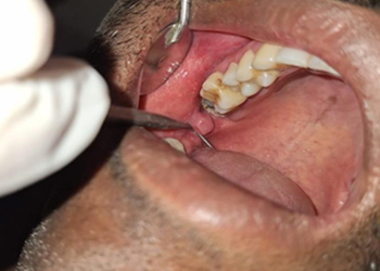
A male patient, 36-year-old, arrived at the Oral Medicine and Radiology Department complaining primarily of a growth that had been present for the previous two months in the area of his right upper back teeth. The patient was apparently normal before 2 months and had no significant dental history. Medical history shows that the patient is overall healthy. The etiology is trauma caused by sharp edged tooth surface of 18.
Intra-oral Examination: On inspection, a single moveable pedunculated growth is detected on the right buccal mucosa, measuring about 1.5X1.5mm and extending 1cm short of the upper mucobuccal sulcus and inferiorly away from the right lower buccal sulcus. The surface above the growth seems smooth. On palpation, the site's size, shape, and extent are confirmed; it is non-tender, with no pus discharge; it has a soft to firm consistency; and it does not bleed when manipulated.([Figure 2])


|
Case |
Age |
Gender |
Site |
Personal history |
Description |
|
|
Case 1 |
59 |
Male |
Labial Mucosa |
Pt has history of smoking from past 4 years. |
On inspection |
On palpation |
|
A single sessile growth on the labial mucosa measuring about 0.5X0.5 and extending from the lower labial mucosa 1.5 mm away to the vermillion border surface is seen. |
On palpation it is soft to firm in consistency,it is compressable , movable and does not bleed when manipulated. |
|||||
|
Case 2 |
36 |
Male |
Buccal mucosa |
No relevant history. |
A single moveable pedunculated growth is detected on the right buccalmucosa, measuring about 1.5X1.5mm and extending 1cm short of the upper mucobuccal sulcus and inferiorly away from the right lower buccal sulcus is seen. |
On palpation it is non-tender, with no pus discharge; it has a soft to firm consistency; and it does not bleed when manipulated. |
|
Case 3 |
45 |
Male |
Buccal mucosa |
No relevant history. |
A single moveable growth on the right buccal mucosa, about measuring 2X2mm, extends 1cm short of the upper mucobuccal sulcus and inferiorly away from the right upper buccalsulcus is seen. |
On palpation it is non-tender, with no pus discharge; it has a soft to firm consistency; and it does not bleed when manipulated. |
Case 3
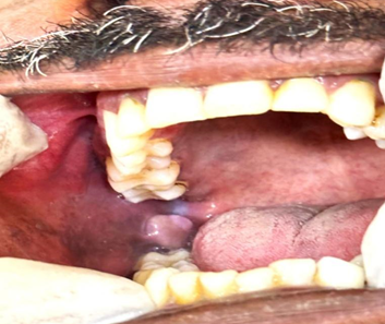
A male patient, 45 years old, at the Oral Medicine and Radiology Department with the primary concern of growth in the area around his right lower back tooth for past four months. The patient was apparently normal before four months and had no significant dental history. Medical history shows that the patient is overall healthy. The etiology is trauma caused by sharp edged tooth surface of 47. Intra-oral Examination: On Inspection A single moveable growth on the right buccal mucosa, about measuring 2X2mm, extends 1cm short of the upper mucobuccal sulcus and inferiorly away from the right upper buccal sulcus; the surface above the growth seems smooth. On palpation, the site, size, shape, and extent are confirmed; it is non-tender, with no pus discharge; it has a soft to firm consistency; and it does not bleed when manipulated.([Figure 3])
Discussion
The term "irritational fibroma" was coined by Dr. E. A. McCarthy. The most prevalent type of connective tissue tumor in the oral cavity is fibroma. A fibrous or granular tissue's inflammation is referred to as "inflammatory hyperplasia". This term describes a benign fibrous growth that typically arises in response to chronic irritation or trauma.Depending on how much the lesion has undergone excessive healing and inflammatory reactions, proliferative benign connective tissue tumors can range in size from tiny to enormous. "Epulis" is the term for a comparable gingival lesion. Localized fibrous hyperplasia, fibromatous fibroma, and irritational fibroma are other names for fibroma. In the third, fourth, and fifth decades of life, fibroma is more common in females than in males. If this benign lesion undergoes malignancy there is increased risk of oral cancer and Malignant lesions can grow rapidly and invade surrounding tissues, potentially causing local tissue destruction, pain, and difficulty with normal oral functions such as eating and speaking. Malignant lesions might have a higher likelihood of recurrence, necessitating ongoing surveillance and possibly multiple treatments. If the malignancy affects critical areas of the oral cavity or jaw, it can impact essential functions like chewing, swallowing, and speaking. [8]
Conclusion
To summarise, irritational fibromas are a common occurrence, often caused by mechanical irritation or persistent damage. While they are typically innocuous, quick identification is critical to alleviating symptoms and avoiding consequences. The buccal mucosa is the most prevalent place for irritational fibroma to develop.[9] Regular dental check-ups can help with early diagnosis and management of these lesions, guaranteeing good oral health.
Source of Funding
None.
Conflict of Interest
None.
References
- J Tomes. A Course of Lectures on Dental Physiology and Surgery, delivered at the Middlesex Hospital School. . Am J Dental Sci 1848. [Google Scholar]
- A Dhanai, H S Bagde, R Gera, K Mukherjee, C Ghildiyal, H Yadav. Case report on irritational fibroma. J Pharm Bioall Sci 2024. [Google Scholar]
- LK Mathur, AP Bhalodi, B Manohar, A Bhatia, N Rai, A Mathur. Focal fibrous hyperplasia: a case report. Int J Dent Clin 2010. [Google Scholar]
- N O Nartey, H A Mosadomr, M Al-Cailani, A Almobeerik. Localized inflammatory hyperplasia of the oral cavity: clinico-pathological study of 164 cases. Saudi Dent J 1994. [Google Scholar]
- C Kar, P Sarkar, S Das, A Ghosh. Large irritation fibroma of palate - a rare presentation. J Pak Assoc Dermatol 2015. [Google Scholar]
- NO Nartey, HA Mosadomr, M Al-Cailani, A Almobeerik. Localized inflammatory hyperplasia of the oral cavity: clinico-pathological study of 164 cases. Saudi Dent J 1994. [Google Scholar]
- IK Salim, AR Diajil. Oral findings, salivary creatinine and urea levels in CKD patients on hemodialysis and on conservative treatment. J Pharm Sci Res 2018. [Google Scholar]
- VA Sangle, VK Pooja, A Holani, N Shah, M Chaudhary, S Khanapure. Reactive hyperplastic lesions of the oral cavity: a retrospective survey study and literature review. Indian J Dent Res 2018. [Google Scholar]
- B Fatani, A I Alhilal, F A Alghamdi, N A Alfawaz, M A Alhaqbani, F S Almutairi, H S Alrfydan. Irritational fibroma mimicking an odontogenic infection: a case report of a misdiagnosed extraoral fibroma. Cureus 2024. [Google Scholar]
