- Visibility 148 Views
- Downloads 25 Downloads
- Permissions
- DOI 10.18231/j.johs.2024.029
-
CrossMark
- Citation
Enhancing smile in class II division 2 malocclusion using utility arch – A Case report
Abstract
Class II Division 2 malocclusion is a dental condition characterized by retroclined maxillary central incisors, often accompanied by a deep bite and a Class II skeletal relationship. The utility arch, an orthodontic appliance designed to exert controlled forces on the dentition, offers a promising approach for correcting this type of malocclusion. By making strategic adjustments, such as a V bend in the vestibular bridge, the utility arch can be effectively used to procline incisors. This case report details the application of a utility arch in treating a growing patient with Class II Division 2 malocclusion. The treatment plan aimed to leverage the patient's growth period to enhance the stability of the correction, ensuring a favorable long-term outcome.
Introduction
Class II Division 2 malocclusion is a specific dental anomaly characterized by a distinct arrangement of the upper and lower teeth. In this condition, the upper first molars are positioned more anteriorly compared to the lower first molars, similar to Class II Division 1 malocclusion. However, a key differentiating feature of Class II Division 2 is the retroclination of the upper central incisors, often accompanied by the proclination of the upper lateral incisors.[1]
This type of malocclusion not only affects the aesthetics of the smile but also has functional implications. Patients may experience difficulties with chewing and speech, as well as an increased risk of temporomandibular joint (TMJ) disorders.[2] The etiology of Class II Division 2 malocclusion is multifactorial, involving genetic predisposition, environmental influences, and habitual factors such as thumb sucking or abnormal tongue posture.
The diagnosis and treatment of Class II Division 2 malocclusion require a thorough understanding of dental and skeletal relationships, as well as an individualized approach to orthodontic therapy. Early intervention can often mitigate the severity of the malocclusion and reduce the complexity of future treatment needs.[1], [2] By addressing both the dental and skeletal components, orthodontists can improve the functional and aesthetic outcomes for patients with this type of malocclusion.
Case Report
A 14-year-old female patient reported to the Department of Orthodontics with the chief complaint of irregularly placed upper and lower front teeth. No contributory medical or dental history.
|
1 |
Morley’s ratio |
100% |
|
2 |
Smile index |
Average |
|
3 |
Smile line |
Average |
|
4 |
Smile arc |
Non- Consonant |
|
5 |
Buccal corridors |
Average |
Extraoral examination ([Figure 1])
Apparently symmetrical
Mesoprosopic face
Convex profile
Competent lips

TMJ Examination: Normal mouth opening, no tenderness on palpation, Normal functional movement with no deviation or deflection with the presence of clicking bilaterally.
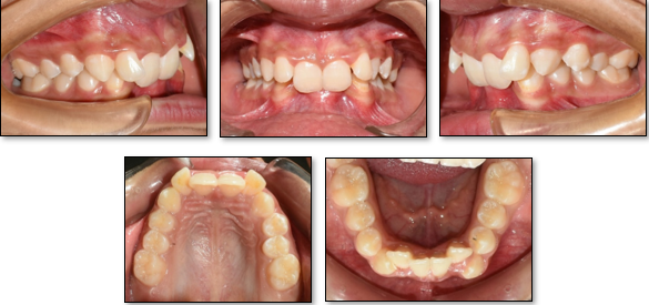
Intraoral examination ([Figure 2])
Class I molar and canine relation bilaterally
Retroclined 11 and 21
Labially tipped 22
Scissor bite in relation to 24 and 34
Traumatic deep bite
Reduced overjet
Overbite – 100%
Freeway space – 5 mm
Crowding in relation to lower anterior teeth
Gingival recession – 31,41
Radiographic examination
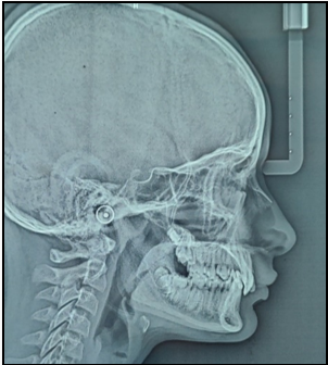
Lateral cephalogram analysis revealed presence of Class II skeletal base, Hypodivergent growth pattern and CVMI – III. ([Figure 3])
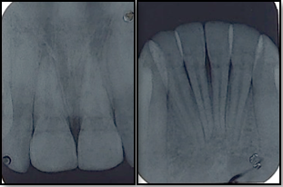
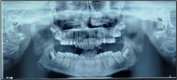
OPG and IOPA examination revealed presence of bone loss in the lower anterior region. ([Figure 4], [Figure 5])
|
S. NO |
Parameters |
Pretreatment |
Posttreatment |
|
Hard Tissue Findings |
|||
|
1 |
SNA |
84˚ |
83˚ |
|
2 |
SNB |
80˚ |
82˚ |
|
3 |
ANB (3.12˚ + 1.8) |
4˚ |
1˚ |
|
4 |
Wits (0 mm) |
5.8 mm |
0 mm |
|
5 |
APP-BPP (5mm) |
5.8 mm |
3.52 mm |
|
6 |
MM Bisector (-5 mm) |
-3.5 mm |
-4.7 mm |
|
7 |
SN length (68.4 mm) |
68.2 mm |
68.2 mm |
|
8 |
Maxillary length (48.9 mm) |
45.8 mm |
44.7 mm |
|
9 |
Mandibular length (75.5 mm) |
71 mm |
74 mm |
|
10 |
FMA (23.83˚ + 2) |
19˚ |
19˚ |
|
11 |
SN-MP (32˚ -35˚) |
23˚ |
24˚ |
|
12 |
Y axis (59.62˚ + 3) |
56˚ |
54˚ |
|
13 |
Bjork sum (394˚) |
128˚ +137˚ +117˚ =382˚ |
128˚ +138˚ +118˚ =383˚ |
|
14 |
Gonial angle (123˚ +7) |
117˚ |
118˚ |
|
15 |
Upper anterior facial height (45%) |
50% |
48% |
|
16 |
Lower anterior facial height (55%) |
50% |
52% |
|
17 |
Ramal height |
44.7 mm |
48.2 mm |
|
Dento-Alveolar Findings |
|||
|
1 |
Mx 1 to A-Pg (6.74 + 1.3mm) |
0 mm |
5.8 mm |
|
2 |
Mx 1 to NA (4.92 + 2.05 mm) |
-3.52 mm |
5.8 mm |
|
3 |
Mx 1 to NA (24.02˚ + 5.82) |
3˚ |
28˚ |
|
4 |
Mx 1 to Palatal plane (71˚) |
91˚ |
62˚ |
|
5 |
Md 1 to A-Pg (-2 to +2 mm) |
-7.05 mm |
1.7 mm |
|
6 |
Md 1 to NB (6 + 1.7 mm) |
-3.52 mm |
4.1 mm |
|
7 |
Md 1 to NB (27˚ + 4.3) |
0˚ |
28˚ |
|
8 |
IMPA (96˚) |
80˚ |
99˚ |
|
9 |
Inter-incisor Angle (123˚) |
180˚ |
122˚ |
|
Soft Tissue Findings |
|||
|
1 |
E – line - Upper lip (-4 mm) |
-2 mm |
0 mm |
|
|
E – line - Lower lip (-2 mm) |
-2.35 mm |
1.1 mm |
|
2 |
S-line - Upper lip (0 mm) |
1.172 mm |
1.1 mm |
|
|
S-line - Lower lip (0 mm) |
0 mm |
1.1 mm |
Study model analysis
Study model analysis revealed discrepancy of 2mm in maxillary arch and 3 mm in mandibular arch with mandibular anterior excess of 1.6 mm.
Problem list
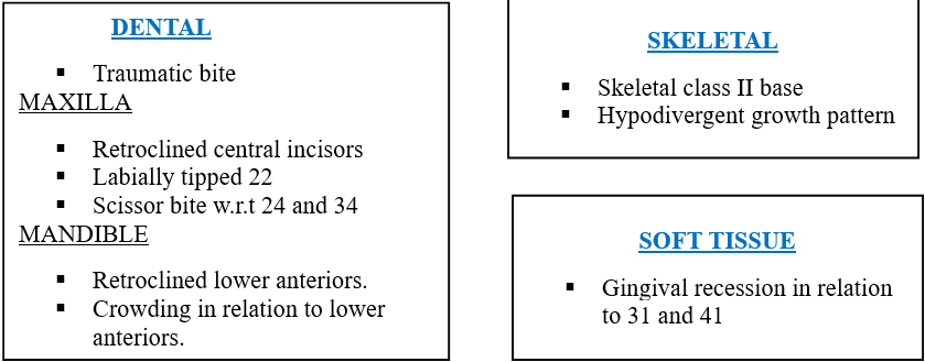
Treatment plan
Utility arch for the correction of retroclined upper incisors followed by fixed orthodontic mechanotherapy with preadjusted edgewise (MBT-022X028” slot) appliance.
Treatment progress
Bonding of upper and lower arch was done. Anterior bite plane was used for the correction of deep bite. Utility arch was given for the correction of retroclined incisors. ([Figure 7])
After the correction of retroclined incisors, alignment and levelling of arches was done, with the progression of wire at every 3-week interval. ([Figure 8])
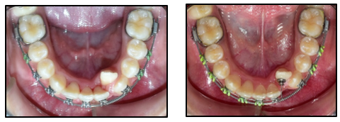
In the mandibular arch, open coil spring was placed in 17x25 inch stainless steel wire to create space for lingually placed 32. ([Figure 9])
Piggy back technique was used for the alignment of 32 in the lower arch ([Figure 10])
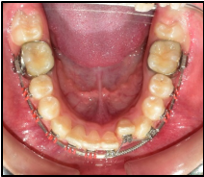
Finishing and detailing of upper and lower arches. ([Figure 11])
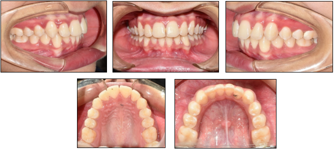
Debonding done ([Figure 12], [Figure 13]) and retainers were delivered. Removable Hawley’s retainer with anterior bite plane in the upper arch and fixed retainer in the lower arch.




Discussion
Class II division 2 malocclusion is a distinct category of class II malocclusion with unique features of retroclined incisors and a strong pattern of familial inheritance.[3] Most Class II Division 2 malocclusions are typically due to a skeletal class II jaw relationship. However, some authors propose that many of these cases actually exhibit a skeletal class I jaw relationship. Patients often present with a hypodivergent facial pattern, short squarish face, with strong musculature.
Although the etiology of Class II Division 2 malocclusion is not well understood, several theories have been proposed. The Class II Division 2 pattern is known to frequently occur within families. Ruf and Pancherz, however, described a case of two monozygotic twins with distinct malocclusions: one had a Class II Division 2 malocclusion and the other a Class II Division 1 malocclusion. They conjectured that there are other etiological factors besides heredity that contribute to Class II Division 2 malocclusion in light of this observation.[4] Some authors suggest that this malocclusion stems from insufficient mandibular development or the mandible being positioned distally relative to the cranial base. Others believe the primary cause is dentoalveolar rather than skeletal in origin. Some clinicians have proposed that the lips act as a local epigenetic factor in Class II Division 2 malocclusion, with maxillary incisor retroclination resulting from excessive, pressure between the lip and teeth.[5]
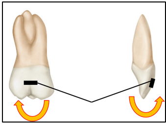
In the present case, a utility arch was employed to correct retroclined incisors. Activation of the utility arch involved creating a V bend in the center of the vestibular bridge. This central V bend generates equal moments at both ends of the arch, leading to the proclination of the retroclined incisors without causing any vertical changes, such as intrusion or extrusion. ([Figure 15])
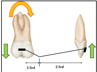
When the V bend is positioned at one-third the distance ([Figure 16]) from the posterior end of the bridge, it results in pure intrusion of the anterior teeth, a clockwise moment, and extrusion at the molars. However, since the patient did not have a gummy smile, intrusion of the incisors was unnecessary, so the V bend was placed in the middle of the bridge. The patient displayed increase in freeway space with no gummy smile, hence the deep bite correction was achieved through the extrusion of the posterior teeth using an anterior bite plate. [6], [7]
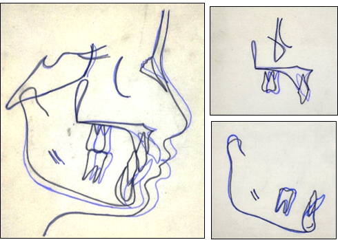
In the present case, the skeletal class II base was corrected with the normal growth of mandible ([Figure 14]) after eliminating the restraining effect of retroclined incisors on the mandible. ([Table 1]) The bone loss and gingival recession caused due to traumatic bite improved following treatment.
Regarding the stability of deep bite correction, it is generally challenging to maintain in hypodivergent patients. However, in this case, since the treatment was carried out during the growth period, the stability of the deep bite correction is expected to be good. Moreover, establishing a proper interincisal angle and fully correcting the curve of Spee will further enhance the stability of the deep bite correction.
Conclusion
In conclusion, the case report demonstrates that the use of a utility arch is an effective and efficient method for enhancing the smile in patients with Class II Division 2 malocclusion. By carefully controlling the proclination of the maxillary incisors and addressing deep bite issues, the utility arch plays a crucial role in improving both the function and aesthetics of the smile.
Source of Funding
None.
Conflict of Interest
None.
References
- William D, Fields H, Larson D, B. . Contemporary Orthodontics. 2019. [Google Scholar]
- Graber L, Vanarsdall R, Vig K, Huang G. . Orthodontics - E-Book: Current Principles and Techniques. 2016. [Google Scholar]
- Kharbanda O. . Diagnosis and Management of Malocclusion and Dentofacial Deformities, E-Book. 2019. [Google Scholar]
- Ruf S, Pancherz H. Class II division 2 malocclusion: genetics or environment? A case report of monozygotic twins. Angle Orthod. 1999;69(4):321-4. [Google Scholar]
- Jain N, Soni S. An overview of class II division 2 malocclusion. Int J Health Sc. 2021;5(S2):214-21. [Google Scholar]
- Choy K. . Burstone’s Biomechanical Foundation of Clinical Orthodontics. 2022. [Google Scholar]
- Valarelli F, Carniel R, Cotrin-Silva P, Patel M, Cançado R, Freitas K. Treatment of a Class II Malocclusion with Deep Overbite in an Adult Patient Using Intermaxillary Elastics and Spee Curve Controlling with Reverse and Accentuated Archwires. Contemp Clin Dent. 2017;8(4):672-8. [Google Scholar]
How to Cite This Article
Vancouver
Aiswareya G, Verma SK, Sharma VK, Yadav PK. Enhancing smile in class II division 2 malocclusion using utility arch – A Case report [Internet]. J Orofac Health Sci. 2024 [cited 2025 Sep 17];11(3):146-151. Available from: https://doi.org/10.18231/j.johs.2024.029
APA
Aiswareya, G., Verma, S. K., Sharma, V. K., Yadav, P. K. (2024). Enhancing smile in class II division 2 malocclusion using utility arch – A Case report. J Orofac Health Sci, 11(3), 146-151. https://doi.org/10.18231/j.johs.2024.029
MLA
Aiswareya, G, Verma, Sanjeev Kumar, Sharma, Vivek Kumar, Yadav, Pramod Kumar. "Enhancing smile in class II division 2 malocclusion using utility arch – A Case report." J Orofac Health Sci, vol. 11, no. 3, 2024, pp. 146-151. https://doi.org/10.18231/j.johs.2024.029
Chicago
Aiswareya, G., Verma, S. K., Sharma, V. K., Yadav, P. K.. "Enhancing smile in class II division 2 malocclusion using utility arch – A Case report." J Orofac Health Sci 11, no. 3 (2024): 146-151. https://doi.org/10.18231/j.johs.2024.029
