Introduction
Dental radiography provides essential information in the diagnosis of oro-facial pathology and subsequent treatment planning. 1 The temporo-mandibular joint (TMJ) imaging is very challenging because the bony components are small and superimpositions from the base of the skull often result in a lack of clear delineation of the joint.2 Although widely used, 2D images, like orthopantomograms (OPG) have a few inherent limitations, namely, magnification and minification of structures, superimposition of anatomical and/or pathological entities, and misrepresentation of structures.3 However, due to its three-dimensional complexity, the TMJ cannot be accurately evaluated by conventional radiographic methods, in all three dimensions, images that overlap. Currently, cone-beam computed tomography (CBCT) is the imaging modality of choice to investigate bone alterations of the TMJ, since it requires less X-ray exposure and lower cost to the patient, than the spiral computed tomography (CT), and it is possible to obtain sections of any structure on several levels, with minimal distortion. 2, 3, 4, 5
Practically, it isn’t viable to assess condylar changes in patients with temporomandibular joint disorders (TMD’s) with multiple imaging modalities, owing to high radiation and high imaging costs. Moreover, anthropological and forensic studies rely heavily on the documentation of morphological variations of the TMJ components. In forensic dentistry, radiographs play a crucial role in revealing details that would otherwise go undetected on visual inspection alone. Therefore, the present study aims to assess the validity of OPG, by evaluating the same subjects on OPG and CBCT and comparing the morphological variations in the shape of condyle, coronoid process and sigmoid notch.
Materials and Methods
The present retrospective study was conducted in the department of oral medicine and radiology, after obtaining the ethical approval from the institutional ethical committee and obtaining an informed consent from the subjects at the time of acquisition of scan, to use and share the information from the scan for purposes of education, including teaching and research publications, as per the protocol of our institution. Sample size of 200 was calculated. For the transparency of the study reporting, the Strengthening the Reporting of Observational studies in Epidemiology (STROBE) guidelines were followed.
All clinically justified scans, showing TMJ completely with the demographic data, indication of scan or referral note from the other department will be included in the study. Images of subjects with previous history of surgery or trauma, and with poor image quality or any artefacts will be excluded from the study.
The images were acquired using GENORAY PAPAYA 3D PLUS Combination Imaging System (Genoray Co., Ltd., South Korea) with Triana imaging software. The sensor type was thin film transistor (TFT), scan mode was continuous for OPG and pulsed for CBCT, exposure parameters range from 60-90 kVp and 4 – 12 mA, field of view and time for acquisition of the scans for OPG was 75 x 75 µ and 9-17 seconds and for CBCT was 16 x 14 and 7.7-14.5 seconds. The software used to analyze the scans was TRIANA-Genoray’s 3D reconstruction viewer version and the reading of scans was done using a 24” HP workstation flat-screen LCD monitor, with 1080 pixel resolution and maximum color quality (64 bits).
After following the inclusion and exclusion criteria, 100 OPG and CBCT scans were included in this study, i.e. a total of 200 temporomandibular joints. The demographic data for each subject was recorded. Then positional adjustment of the multi-dimensional images on the monitor screen was done and examination of each OPG and CBCT scan was conducted in coronal, axial, and sagittal planes, in a systematic fashion.
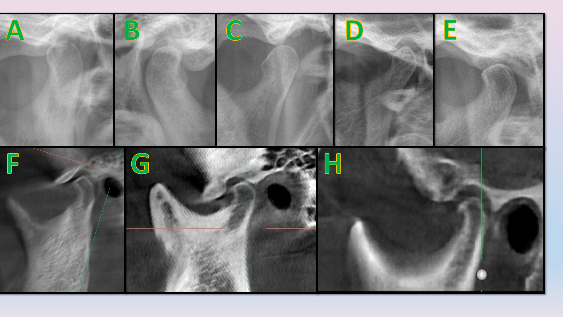
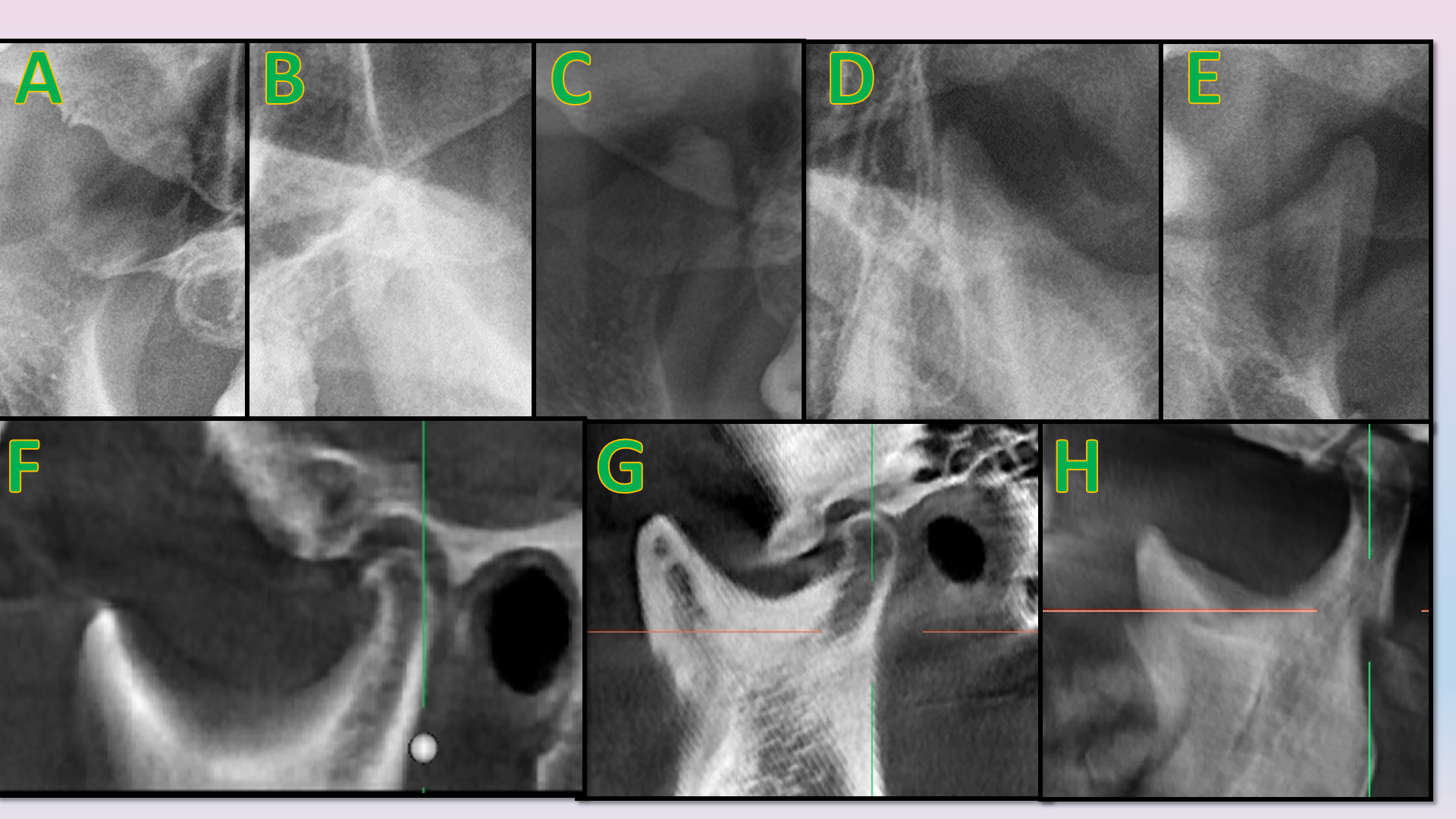
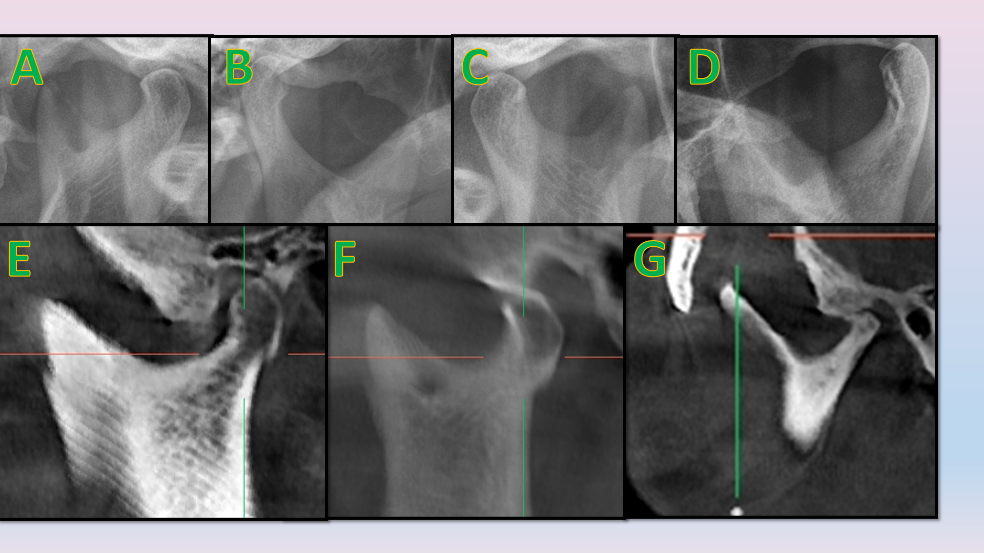
Anatomical variations of condyle on OPG (Figure 1A, 1B, 1C, 1D, 1E) were classified into round, bird beak, crooked finger, diamond and flat, based on the classification provided by Mahapatra et al.6 whereas, anatomical variations of condyle on CBCT (Figure 1F, 1G, 1H) were classified into round, flat, beak and concave, based on the classification provided by Koyama et.al.7 and Shubhasini et al. 2 Anatomical variations of coronoid process on OPG (Figure 2A, 2B, 2C, 2D, 2E) were classified into round, triangle, crooked finger, flat and beak, based on the classification provided by Mahapatra et.al. 6 whereas, anatomical variations of coronoid process on CBCT (Figure 2F, 2G, 2H) were classified into triangular, round and beak/hook, based on the classification provided by Nayak et.al.8 Anatomical variations of sigmoid notch on OPG (Figure 3A, 3B, 3C, 3D) were classified into round, sloping, V-shaped and flat, based on the classification provided by Mahapatra et.al.6 whereas, anatomical variations of sigmoid notch on CBCT (Figure 3E, 3F, 3G) were classified into sloping, wide and round, based on the classification provided by Shakya et.al.9
Anatomic variations were detected by well-established radiographic interpretation process and diagnostic criteria, and all of the findings were noted on the proforma designed for this study. Statistical analysis was performed.
Figure 1
Anatomic variations of condyle on OPG (A = Round, B = Crooked finger, C = Bird beak, D = Diamond, E = Flat) and sagittal section of CBCT (F = Flat, G = Round, H = Beak)

Figure 2
Anatomic variations of coronoid process on OPG (A = Round, B = Beak, C = Crooked finger, D = Triangular, E = Flat) and sagittal section of CBCT (F = Triangular, G = Round, H = Beak/hook)

Figure 3
Anatomic variations of sigmoid notch on OPG (A = Round, B = Sloping, C = Flat, D = V-shaped) and sagittal section of CBCT (E = Wide, F = Round, G = Sloping)

Table 1
Gender-wise and Age-wise distribution of subjects
|
Demographics |
Subjects (n=100) |
Total |
|
|
Gender |
Male |
60 (60%) |
100 (100%) |
|
Female |
40 (40%) |
||
|
Age Group |
21-40 years |
28 (28%) |
100 (100%) |
|
41-60 years |
42 (42%) |
||
|
61 years and above |
30 (30%) |
||
Table 2
Gender-wise distribution of anatomic variations of shape of condyle, coronoid process and sigmoid notch on OPG
Table 3
Gender-wise distribution of anatomic variations of shape of condyle, coronoid process and sigmoid notch on CBCT
Table 4
Age-wise distribution of anatomic variations of shape of condyle, coronoid process and sigmoid notch on OPG
Table 5
Age-wise distribution of anatomic variations of shape of condyle, coronoid process and sigmoid notch on CBCT
Results
Out of 100 scans analyzed, 60 were of males (60%) and 40 were of females (40%). [Table 1] 28 (28%) belonged to the age group of 21-40 years, 42 (42%) belonged to the age group of 41-60 years and 30 (30%) belong to the age group of 61 years and above. [Table 1] The most common shape of condyle, coronoid process and sigmoid notch on OPG was round (30%), round (40%) and round (40%) respectively, whereas on CBCT the most common shape of condyle, coronoid process and sigmoid notch were flat (58%), triangular (34%) and wide (37%) respectively. [Table 2 & 3] The sensitivity and specificity of OPG was found to be 33% and 68% respectively.
Discussion
The function and health of TMJ is vital to life. The functions of the TMJ are to provide smooth, efficient movement of the mandible during mastication, swallowing, and speech and to provide stability of mandibular position and prevent dislocation from external or unusual forces. A normal variation in the morphology of bony components of TMJ may occur with age and different genders, facial types, occlusal forces etc. 10 Morphological variations depend upon developmental variation along with condylar remodelling to accommodate malocclusion, trauma, infections and other pathological and developmental abnormalities such as tumors, condylar or coronoid hyperplasia, ankylosis etc. 11, 12 Variability in the shapes and sizes of condyle, coronoid and sigmoid notch helps to diagnose the TMJ disorders associated with malocclusions such as crossbite, deep bite, and open bite. 12 These factors can alter dynamic functionality (opening and closing) or trigger problems such as occlusal instability, articular clicks, joint and muscle pains, TMJ alterations (resorption), functional problems, mandibular deviations and dislocations. 11 The abovementioned morphological variations may be indispensable aids in anthropological and forensic studies and therefore, it is necessary to assess the prevalence and document various anatomic variations in the shapes of condyle, coronoid and sigmoid notch.
The most common shape of condyle on OPG in the present study was round (30%), whereas the least common shape was flat (10%), which is highly significant (p = 0.000**). This is similar to the results of the study conducted by Sahithi D et.al. 13 who also reported that the common shape of condyle on OPG was round (39%), whereas the least common shape of condyle was flat (4%). 13
The most common shape of condyle on OPG in males was crooked finger, whereas the least common shape was flat. However, the most and least common shapes of condyle on OPG in females were round and flat respectively. These results are in accordance with the results of the study conducted by Mahapatra et.al. 6 who also reported that the most common condylar shape in males and females was round and the least common was flat. Moreover, the abovementioned findings are similar to those reported by Gupta et.al. 14 who also reported the most common condylar shape in females to be round. However, they reported the least common shape in females was crooked finger, whereas the most common shape of condyle in males was round and the least common shape was crooked finger. These differences could be attributed to a large sample size of the study conducted by Gupta et.al. 14 and their inclusion of all morphological variations present in the literature. When the anatomic variations in shape of condyle were correlated with age, we found that the most common shape in the 21-40 years, 41-60 years and > 60 years were round, round and crooked finger respectively. Similar to our results, Shaikh AH et.al. 12 also reported that oval shape of condyle was predominant in younger age groups, however, their results showed that in older age group too, oval was the most common shape of condyle. This difference could be due to variations in sample size and categorizing age groups in their study and the present study.
However, on CBCT, the most common condylar shape in sagittal section was flat (58%), with the most common shape in males being flat, whereas in females it was round. This is similar to the findings of Tassoker M et.al. 15 who reported that the most common condylar shape on CBCT in sagittal section was in females was round, however, in contrast to the present study, they reported that in males, the most common shape was round. Moreover, when the condylar shapes in sagittal section on CBCT were correlated with age groups, the most common shape in all the age groups was flat. This is similar to the results of Tassoker M et.al. 15 who also reported the most common condylar shape in sagittal section in adults and old age groups to be flat, however, they reported the most common shape to be round, in the young population. The abovementioned differences could be attributed to a different male: female ratio (0.7:1) in their study, compared to the present study (1.5:1).
The most common shape of coronoid process on OPG in the present study was round (40%), whereas the least common shape was flat (4%), which is highly significant (p = 0.007**). This is similar to the results of the study conducted by Sahithi D et.al. 13 who also reported that the least common shape of coronoid process was flat (2%), however, in their study the most common shape of coronoid process was triangular (54%). This slight discrepancy could be due to a larger sample size and a different classification of morphological variations used by the abovementioned author, compared to the present study. The most and least common shapes of coronoid process on OPG in males were round and flat respectively. However, the most and least common shapes of condyle on OPG in females were beak and flat respectively. These results are comparable to those reported by Mahapatra et.al. 6 who also reported that the most common shape of coronoid process in males was round. However, a dissimilarity was noted between their results and those of the present study, since they reported that beak shape was the lease common shape of coronoid process in males and the most and least common shapes of coronoid process in females was round and beak respectively. These differences could be because they had a large sample size. When the shape of coronoid process on OPG was correlated with age, the most common shape in the 21-40 years, 41-60 years and > 60 years were round, beak and round respectively. These results vary from those of Bains SK et.al. 16 who reported that the most common shape of coronoid process in all age groups was triangular and the least common was beak, except in the age group of 40-59 years, where the least common shape of coronoid process was flat. These differences could be due to a difference in the sample size, geographic location of population and the classification of morphological variations between the abovementioned and the present study.
However, on CBCT, the most common shape of coronoid process in sagittal section was triangular (34%), with the most common shape in males being triangular, whereas in females it was round. These results are similar to the findings of Tassoker M et.al. 15 who also reported that the most common shape of coronoid process in males on CBCT was triangular, nevertheless, their results showed that in females also the most common shape of coronoid process was triangular. Moreover, when the shape of coronoid process on CBCT was correlated with age groups, the most common shape in the 21-40 years, 41-60 years and > 60 years were round, triangular and beak/hook respectively. However, Tassoker M et.al. 15 reported that the most common shape of coronoid process on CBCT was triangular. The abovementioned differences could be attributed to a different male: female ratio and the fact that their study was conducted in a different population, which could have led to these endemic differences. Also, they classified shapes of coronoid process into triangular and round only, whereas the present study classified it into 3 shapes.
The most common shape of sigmoid notch on OPG in the present study was round (40%), whereas the least common shape was V-shaped (10%), which is highly significant (p = 0.006**). These results are dissimilar to the study conducted by Sahithi D et.al. 13 who reported that the most common shape of sigmoid notch was wide (44%) whereas the least common was sloping (23%). These differences could be attributed to the fact that their study had a larger sample size and it was conducted in South India, whereas the present study had a smaller sample size and was conducted in the western region of India, which could account for ethnic variations in the normal anatomy. The most and least common shape of sigmoid notch on OPG in males were round and V-shaped respectively. However, the most and least common shapes of sigmoid notch on OPG in females were sloping and V-shaped respectively. These results are similar to those reported by Mahapatra et.al. 6 who also reported that the most common shape of sigmoid notch in males was round, and the least common shape in males and females was V-shaped. However, according to their results, the most common shape in females was round. This variation in results from that of the present study could be attributed to a larger sample size of their study. When the shape of sigmoid notch on OPG was correlated with age, the most common shape in the 21-40 years, 41-60 years and > 60 years were round, round and flat respectively, whereas the least common was amongst all age groups was V-shaped. These results are in contrast to those reported by Bains SK et al. 16 who reported that the most common shape of sigmoid notch was wide, with round being the most common shape, with a similar prevalence of wide, only in the age group of 20-29 years. Moreover, they reported that the least common shape in all age groups was sloping. Again, these differences could be due to a difference in the sample size, geographic location of population and the classification of morphological variations between the abovementioned and the present study.
However, on CBCT, the most common condylar shape in sagittal section was wide (46%), with the most common shape in males being wide, whereas in females it was round. These results are similar to the findings of Tassoker M et.al. 15 who also reported that the most common shape of sigmoid notch in females on CBCT was round, but their results showed that in males also the most common shape of sigmoid notch was round. Moreover, when the shape of coronoid process on CBCT was correlated with age groups, the most common shape in the 21-40 years, 41-60 years and > 60 years were round, wide and wide respectively. However, Tassoker M et.al. 15 reported that the most common shape of coronoid process amongst all age groups on CBCT was round. The abovementioned differences could be due to the fact that their study was conducted in the Turkish population whereas the present study was conducted in the Indian population, amounting for racial diversities.
The sensitivity and specificity of OPG was found to be 33% and 68% respectively. This is comparable to the results of Hegde V et.al. 17 who reported the sensitivity and specificity of OPG to be 24.14% and 100% respectively. The slight difference obtained could be because their study was conducted in-vitro, using dry human skulls and artificially created lesions on the condyles.
Conclusion
Morphologic variation in condyle, coronoid process and sigmoid notch occur based on age, gender and racial diversities. Nonetheless, these may be affected due to harmful oral habits such as bruxism, unilateral chewing, trauma, etc. Since OPG is cost-effective and widely available and used, attention needs to be drawn on anatomic variation of TMJ on OPG, which might lead to degenerative diseases of TMJ in the future. Based on the results of the present study, it may be concluded that panoramic radiographs are useful for predicting the morphologic variations of mandibular condyle, coronoid process and sigmoid notch, when compared against the confirmatory radiographic modality like CBCT.
